Romance of Abdomen Best Seen on Which Xray
Answer 1 of 2. If unable to roll patient do cross-table lateral abdomen with.

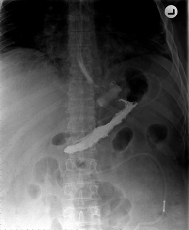
Karenhealey Amazing X Ray Radiology Diagnostic Imaging
Upright AP Projection - Abdomen.
. Abdominal masses air fluid levels and accumulation of intraperitoneal air under the diaphragm is best demonstrated in this position. Abdominal or Abdomen X-rays may be used to help properly place catheters and tubes used for feeding or to decompress organs such as the gallbladder and kidneys. Assess bones and joints on an abdominal X-ray.
An x-ray shows us images because the x-ray passes through different substances at different rates For instance it easily passes through airgas so it is clear whereas it has a hard time with bone and metal so they are dark. The examination should be tailored to each patient. It is also use as a preliminary evaluation radiograph or Scot films for some of special procedure.
X-rays of the pelvis can diagnose arthritis that affects the hip as well as pelvic fractures. Localised or widespread bone disease may give rise to abdominal symptoms. An abdominal X-ray is a safe and painless test that uses a small amount of radiation to make an image of a persons abdomen belly.
An abdominal film is an X-ray of the abdomen. Lower Extremities X Ray Positioning 39 Terms. Radiologists consider an abdomen X-ray to be of good quality when the entire abdomen is visible from the diaphragm superiorly to the pelvis inferiorly.
IPM - Radiography - X-ray parts 45 Terms. Bone and metal show up as white on X-rays. X-ray of abdomen can detect various anomalies in the abdominal cavity.
The abdominal X-ray of this kind is asked for frequently by the doctors though it. Free Download HD or 4K Use all videos for free for your projects. In severe acute abdominal pain plain X-ray of the abdomen is usually the first investigation.
See Chapter 5 for a more complete discussion of abdominal CT protocols. They have their uses for which each is requested. It is best to obtain a general survey examination that includes the entire abdomen and pelvis.
There are many reasons why a doctor may take an abdominal film including to look at organs find infections diagnose pain and look for growths. X-rays of the belly may be done to check the area for causes of abdominal pain. Abdominal X-rays may be taken in the following positions.
Ultrasonography is the better choice in evaluating gallstones. Occasionally there will be unexpected bone disease seen on an abdominal. The AP projection of the abdomen is sometimes called as KUB xray because in this projection the Kidneys Ureters and Bladder are included in the radiograph.
Radiography of upper and lower GI 114 Terms. Also known as the omental bursa is the cavity in the abdomen that is formed by the lesser and greater omentum. AXR typically demonstrates an abnormal bowel gas pattern characterized by dilated small bowel loops with air-fluid levels and a paucity of colonic gas Aka et al 2021.
The selection of an imaging technique depends on the most likely diagnosis clinical setting and local expertise. Bone disease may be incidental or pivotal to diagnosis. Upright xray examination of the abdomen is usually suggested when the Kidneys Uriters and Bladder KUB are not the primary interest to be examine.
An abdominal or pelvic X-ray can help find kidney stones bladder stones and gallstones. In Plain x ray of the abdomen no contrast is used. But the chance of damage from the X-r.
Natural contrast between the kidneys and the low density retroperitoneal fat that surrounds them means they are often visible on an X-ray of the abdomen. If you suspect bone or joint disease then dedicated images of the area in question are required. They lie at the level of T12-L3 and lateral to the psoas muscles.
A systematic approach to the interpretation of abdominal X-rays is described by the mnemonic BBC for bowel and other organs liver gallbladder stomach kidneys spleen bladder bones and calcifications and. On the other hand about 15 of gallstones are radiopaque and are seen on abdominal plain radiograph. Download and use 10000 Hot Bedroom Romance Most Romantic Scene stock videos for free.
It can also be done to find an object that has been swallowed or to look for a blockage or a hole in the intestine. This view is normally performed when localizing foreign bodies or lines within the abdominal cavity. A fold of the peritoneum that attaches the stomach small intestine pancreas spleen and other organs to the posterior wall of the abdomen.
The patient is laying 30 degrees either LAO or RAO often on a 30-degree wedge to ease of positioning. Additionally the oblique abdominal series can be utilized in the assessment of the upper intestinal tract during barium studies. This image shows the stomach liver spleen small and large.
Contrast abdominal x-ray such as in Barium swallow and Barium Meal and Barium Enema requires the use of Barium as the contrast. During the examination an X-ray machine sends a beam of radiation through the abdomen and an image is recorded on special film or a computer. PA Chest UPRIGHT PA or AP Abdomen Include both hemidiaphragms SUPINE Abdomen Include symphysis pubis A LEFT LATERAL DECUBITUS view is to be done on those cases where the patient is unable to stand.
Beaking of the cecal margins with obstruction of the upstream ileum is commonly seen. Free air seen on X-ray of abdomen itself is not threatening. If the patient cannot stand for an erect abdominal film and a PA view of the chest the cross.
Gallstones if they are large and calcified Pancreatic calcifications in pancreatic cyst or chronic pancreatitis Air under the diaphragm in stomach or bowel perforation Air in the bowel in bowel obstruction. Which of the following pathologic conditions is most common in young children. Which two structures create the romance of the abdomen.
For example if you are suspecting stone in kidney X-ray of kidney ureter and bladder makes the diagnosis of the disease easier. The X-ray of the abdomen taken with the patients body in the supine position comprise a large proportion of the plain-film examinations conducting on abdomen. The routine abdominal films consist of supine and upright views.
Only the right diaphragm must be included. X-rays of the abdomen or pelvis are. When an AP Projection of abdomen taken in supine it is also called as Flat Plate Abdomen X-ray.
There are different types of plain x ray films of the abdomen and all have their indications ie. There is always a slight chance of damage to cells or tissue from radiation including the low levels of radiation used for this test.

Abdomen Xray Signs X Ray Bowel Obstruction Abdomen

What Is An Abdominal Or Abdomen Xray Two Views
Abdominal X Ray


Comments
Post a Comment